
Jump to:
What is Pancreas or Pancreatic Cancer?
–Types of Pancreatic Cancer Tumors
Causes
Signs and Symptoms
–Risks of Developing Pancreatic Cancer
Diagnosis
–How is Pancreatic Cancer Diagnosed?
What is Pancreas or Pancreatic Cancer?
 Pancreatic cancer is the fourth most common cause of cancer death in the US.
According to the American Cancer Society’s most recent estimates for pancreatic cancer in the United States, more than:
- 42,000 cases are diagnosed each year
- 35,000 people die because of the disease
The lifetime risk of having pancreatic cancer is about 1 in 72. It is about the same for men and women. The risk increases with age, and most cases are diagnosed in people between 60 and 80 years old.
The pancreas is a spongy, oblong organ about 6 inches long and 2 inches wide. It is located behind the lower part of the stomach, between the stomach and the spine. The pancreas is important because it makes insulin and other hormones that help the body absorb sugar and control blood sugar, and produces juices that aid in digestion.
It is mainly composed of the exocrine cells that make digestive enzymes and the endocrine (islet) cells that make hormones such as insulin, which controls blood sugar levels. Pancreatic cancer occurs when a cell in the pancreas is damaged and starts to grow out of control.
The vast majority of pancreatic cancers start in exocrine cells found in the pancreas ducts, or small channels that carry digestive enzymes to the intestines. These cancers are called adenocarcinomas that begin in the tissue lining the pancreas, although as many as 20 different types of tumors can be found in the pancreas. As a pancreatic tumor grows, it can invade nearby organs – such as the bile duct, intestine, or stomach – or adjacent blood vessels. Tumor cells can break away and spread to the lymph nodes or liver, or to other places in the abdomen. The outcome for individuals with pancreatic cancer depends on the size and type of the tumor, lymph node involvement, and degree of metastases (spreading) at the time of diagnosis.
Dr. Bilchik and his team diagnose and treat pancreatic cancer with the most advanced therapies available. They provide focused, personalized care with a team that consists of specialist surgeons, oncologists, radiologists, gastroenterologists and specially trained support staff, all to help you make the most informed decision about your care.
As a center that has advanced technology and expertise, they offer the most innovative treatments, including minimally invasive surgery, focused radiation therapy, chemotherapy and new immunotherapy drugs.
Types of Pancreatic Cancer Tumors
The pancreas contains two main types of cells:
- Exocrine cells that produce digestive juices
- Endocrine cells that produce hormones
Adenocarcinoma starts in exocrine cells and accounts for 95% of pancreatic cancers. It occurs in the lining of the pancreatic ducts.
Neuroendocrine tumors involves endocrine cells (islet cells). Most of these tumors are malignant, but insulin-producing islet cell tumors are often benign (non-cancerous). Neuroendocrine tumors can be:
- Functional and produce abnormally high amounts of hormones (insulin, gastrin, glucagon) leading to symptoms such as light headedness, pain, weakness, low and high sugar levels and jaundice.
- Non-functional and produce no hormones
Insulinoma is a rare pancreatic tumor that secretes insulin, the hormone that lowers glucose levels in the blood.
Gastrinoma is a tumor that secretes above-average levels of gastrin, a hormone that stimulates the stomach to secrete acids and enzymes. Gastrinoma can also cause stomach ulcers and can spread to the liver.
Glucagonoma is a tumor that secretes glucagon, a hormone that raises levels of glucose in the blood, often leading to a characteristic rash
Other rare pancreatic tumors:
Pancreaticoblastoma is very rare. This type of pancreatic cancer is found mostly in young children. Isolated sarcomas and lymphomas can also occur in the pancreas. These are also very rare.
Pseudopapillary neoplasms are mostly found in women in their teens and 20s. Patients can present with pain, jaundice, weight loss or a mass. These cancers mainly originate in the head of the pancreas. Resection of the tumor is often curative.
Ampullary cancer is a rare type of exocrine tumor that begins where the bile duct from the liver and the pancreatic duct join with the small intestine. Since it causes jaundice, it may be found earlier than other types of pancreatic cancer and therefore has a better outcome.
Causes of Pancreas or Pancreatic Cancer
The cause of pancreatic cancer is largely unknown.
The following conditions are associated with a greater chance of getting pancreatic cancer
- Diabetes
- Smoking
- Obesity
- Alcohol abuse
- Family History
Signs and Symptoms of Pancreas or Pancreatic Cancer
There are no early warning signs for pancreatic cancer. In fact, symptoms may be so non-specific they are often ignored.
The following are the other most common symptoms associated with pancreatic cancer:
- Pain in the upper abdomen (belly) or upper back
- Loss of appetite
- Weight loss
- Jaundice (yellow skin and eyes, and dark urine)
- Indigestion
- Nausea
- Vomiting
- Extreme fatigue
- An enlarged abdomen from a swollen gallbladder
- Pale, greasy stools that float in the toilet
Many of these symptoms can also be caused by other, more common health problems. For example, hepatitis, gallstones, and other liver problems can block the bile duct and are much more common causes of jaundice.
Other signs of pancreatic cancer, such as an enlarged gallbladder or the sudden onset of type II diabetes, may be found by a physician during an exam, even if there are no other noticeable symptoms.
What are the risks of developing pancreatic cancer?
Risk factors for pancreatic cancer include:
- Age
- Most pancreatic cancer occurs in people over the age of 55.
- Smoking
- Heavy cigarette smokers are two or three times more likely than nonsmokers to develop pancreatic cancer.
- Obesity and physical inactivity
- Pancreatic cancer is more common in people who are very overweight and in people who are not physically active.
- Diabetes
- Pancreatic cancer occurs more often in people who have type 2 diabetes than in those who do not.
- Gender
- More men than women are diagnosed with pancreatic cancer.
- Genetic factors
- Approximately 10 percent of people with pancreatic cancer have one or more inherited genetic mutations that can also cause other diseases, including familial atypical multiple mole melanoma syndrome, familial breast cancer, Peutz-Jeghers syndrome, and hereditary pancreatitis.
- Mutations in the genes BRCA1 and BRCA2, which increase the risk of breast, prostate, and certain gynecologic cancers, have been found in some families with a history of pancreatic cancer. Other inherited genetic factors have been identified, but do not greatly increase an individual’s risk of developing pancreatic cancer.
- Race
- African-Americans are more likely than Asians, Hispanics, or whites to be diagnosed with pancreatic cancer.
- Family history
- The risk for developing pancreatic cancer is higher if a person’s mother, father, or a sibling had the disease. An estimated 5 to 10% of people with pancreatic cancer have one or more family members who have had the disease. According to the National Cancer Institute, people with a strong family history of pancreatic cancer are nine times more likely to develop pancreatic cancer than others.
- Cirrhosis of the liver
- People with cirrhosis have a higher risk of pancreatic cancer.
- Workplace exposures
- Exposure to certain occupational pesticides, dyes, and chemicals used in the metal industry may increase the risk of pancreatic cancer.
- Chronic pancreatitis
- Long-term inflammation of the pancreas, often caused by excessive alcohol abuse, has been linked to an increased risk for pancreatic cancer.
- Environmental Factors
- Exposure to carcinogens such as asbestos, pesticides, dyes, and petrochemicals may be linked to pancreatic cancer.
- Benign and Pre-cancerous Pancreatic Lesions
- Advances in imaging technology have dramatically increased the number of small abnormalities that are found in the pancreas. Most of these abnormalities are identified during imaging for another condition. Many of them are benign, fluid-filled cysts and are unlikely to cause symptoms or shorten a person’s life. Others are pre-cancerous and have the ability to turn into pancreatic cancer.
Diagnosis
 One reason for the often poor outcome of pancreatic cancer is the location of the pancreas deep inside the body. Pancreatic tumors can’t be seen or felt by doctors during routine physical exams, and patients usually have no symptoms until the cancer has spread to other organs.
Currently, there are no blood tests to find early cancers of the pancreas. There is a possibility that an extra sensitive probe can be passed into the stomach to better visualize the pancreas (endoscopic ultrasound) and this might be useful in screening people with a high risk of pancreatic cancer.
Blood Tests
A substance called CA 19-9 is released into the blood by exocrine pancreatic cancer cells and can be detected by blood tests. However, by the time CA 19-9 blood levels are high enough to be accurately detected by available methods, the cancer may no longer be in its early stages. This is why the test is not recommended for routine screening of people without symptoms or a known diagnosis of cancer. The CA 19-9 test is sometimes used during treatment to see if the therapy is working or after treatment to see if the cancer has recurred.
Another substance, carcinoembryonic antigen (CEA), can help detect advanced pancreatic cancer in some people. But it isn’t sensitive enough to find the cancer early and is not recommended as a screening test.
Endoscopic Ultrasound
Endoscopic ultrasound (EUS) is a procedure used to image the digestive tract, including the pancreas. A thin, flexible, lighted tube with a small ultrasound probe attached to the end is passed through the patient’s mouth into the stomach and the top part of the small intestine called the duodenum. The ultrasound component of the endoscope uses sound waves to create visual images of the pancreas.
Genetic Testing
Inherited DNA changes are thought to cause as many as 10% of pancreatic cancers. Because these inherited cases are sometimes linked with other cancers, determining whether a patient’s relatives have an increased risk is not simple.
If you’re interested in genetic testing, please talk with Dr. Bilchik and his team who can help interpret and explain the test results and what they may mean.
How is Pancreatic Cancer Diagnosed?
Dr. Bilchik and his team will take a complete medical history and do a physical exam prior to beginning any diagnostic procedures for pancreatic cancer. If pancreatic cancer is suspected, the following tests may be performed in order to make a definitive diagnosis.
Ultrasound (also called sonography)
This diagnostic imaging technique uses high-frequency sound waves to create an image of the internal organs, specifically the liver, pancreas, spleen, and kidneys and to assess blood flow through various vessels. The ultrasound may be done with either an external or internal device:
- Transabdominal ultrasound. The technician places an ultrasound device on the abdomen to create the image of the pancreas.
- Endoscopic ultrasound (EUS). The doctor inserts an endoscope, a small, flexible tube with an ultrasound device at the tip, through the mouth and stomach, and into the small intestine. As the doctor slowly withdraws the endoscope, images of the pancreas and other organs are made.
Your doctor can use these pictures to determine the size and location of a tumor in the pancreas and whether the tumor has spread to nearby blood vessels or other structures. Endoscopic ultrasound can also allow passage of a needle into a suspected tumor to obtain tissue samples. This is a type of biopsy called fine-needle aspiration. Cells obtained from the biopsy can then be analyzed to see if they are cancerous; this information can be used to guide treatment.
Computed Tomography Scan (CT or CAT scan)
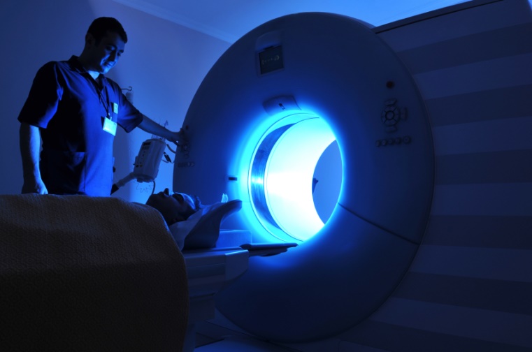 This diagnostic imaging procedure uses a combination of X-rays and computer technology to produce horizontal, or axial, images of the body. A CT scan shows detailed images of any part of the body, including the bones, muscles, fat, and organs. CT scans are more detailed than general X-rays. This is the most important test to look for pancreas cancer and to evaluate whether it can be removed during surgery or whether it has spread to other organs such as the liver or lungs.
Magnetic Resonance Imaging (MRI)
An MRI is a diagnostic procedure that uses a combination of large magnets, radio-frequencies, and a computer to produce detailed images of organs and structures within the body. Specific MRI evaluation of the bile duct, pancreas and pancreatic duct is called magnetic resonance cholangiopancreatography (MRCP).
Endoscopic Retrograde Cholangiopancreatography (ERCP)
This procedure is used to diagnose and treat problems in the liver, gallbladder, bile ducts, and pancreas. A long, flexible, lighted tube called an endoscope is guided through the patient’s mouth and throat, then through the esophagus, stomach, and duodenum (first part of the small intestine). A tube is passed through the scope, and a dye is then injected to allow the bile and pancreatic ducts to be seen on an X-ray.
Percutaneous Transhepatic Cholangiography (PTC)
During a PTC test, a needle is inserted through the skin and into the liver. Dye (contrast) injected through the needle allows the bile duct structures to be seen via X-ray. This test is generally only done if an ERCP cannot be done.
Pancreas Biopsy
A sample of pancreatic tissue is removed with a needle or during surgery and then examined under a microscope.
Special Blood Tests
In addition to the CA 19-9 blood test other blood tests may be necessary to determine if the pancreas tumor is a neuroendocrine type tumor. These include chromogranin, insulin, gastrin, glucagon, serotonin, 5HIAA and pancreastatin.
Positron Emission Tomography (PET)
This is a type of nuclear medicine procedure. For this test, a radioactive substance, usually bound to a type of sugar, is injected through a vein before the body is scanned. Cancer cells take up sugar more than normal cells and this shows up on the x-ray image as a dark spot. This test is not as specific as CT scanning, and is not used alone to diagnose pancreatic cancer. A PET scan is often done in combination with a CT scan and may be useful in evaluating whether cancer has spread to other organs such as the liver or lung.